Components of the Knee
Being the largest (Schmidler, 2016) and one of the most important joints in the human body, the knee joint is composed of several types of tissues and structures. The knee joint is composed of bone, muscle, cartilage, tendons/ligaments, and other specialized connective tissue structures (Martin, Nath, & Bartholomew, 2012). The knee joint itself forms the articulation between the femur of the upper leg and the tibia in the lower leg, and a second articulation between the femur and the patella, the knee cap (Schmidler, 2016).
Typical Knee Joint (taken from: http://orthoinfo.aaos.org/topic.cfm?topic=a00549)
All connective tissue structures of the knee allow for both flexion and extension movements as well as some slight rotation, giving the knee its range of motion (Schmidler, 2016). These connective tissue structures include, but are not limited to, the bones that make up the knee joint (femur, patella, tibia, fibula), hyaline cartilage that protects bone and allows for the sliding movement of the joint, fibrous cartilage of the meniscus which resists pressure placed on the joint, and various ligaments, such as the anterior cruciate ligament, the posterior cruciate ligament, and the medial and lateral collateral ligaments (Schmidler, 2016). Ligaments are strong bands of connective tissue that are not particularly flexible. As such, they are prone to tearing and/or snapping, with these tears making up a large amount of knee injuries (American Academy of Orthopaedic Surgeons, 2014).
Functions of Knee Structures
The structures of the knee allows this joint to bear most of the weight of the body (Schmidler, 2016). When standing, the joint is put into action, with the tibia and femur locking together to form the stable unit that bears the weight of the body.
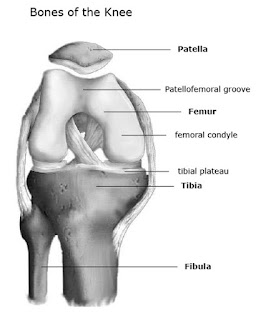
The four bones that make up the knee give strength, stability, and flexibility to the joint (Schmidler, 2016). The functions of the various knee ligaments include attaching the bones of the knee to one another as well as to give strength and stability to the very fragile joint itself. The collateral ligaments (medial and lateral) keep the move from moving too far side to side, and the two cruciate ligaments (anterior and posterior) allow the tibia to move back and forth under the femur. Overall, the four ligaments of the knee are the most vital structures in controlling knee stability and movement (Schmidler, 2016).
Bones of the Knee Joint (taken from: http://www.healthpages.org/anatomy-function/knee-joint-structure-function-problems/)
Cartilage is a type of connective tissue specialized for providing tissues tensile strength and firm structural support (Eroschenko, 2013). The properties of cartilage allows for flexibility without distorting the tissue and makes the tissue resistant to compression (Schmidler, 2016). Specifically within the knee, hyaline cartilage covers the end of the femur, the top of the tibia, and the back of the patella, and fibrous cartilage, in the form of the lateral and medial menisci, act as shock absorbers for the joint (Schmidler, 2016).
Ligaments of the Knee Joint (taken from: http://www.healthpages.org/anatomy-function/knee-joint-structure-function-problems/)
Knee Pathology
Although the many structures of the knee provide great stability, knee injuries are still very common. Sports injuries, specifically, are one of the main causes of these problems (Schmidler, 2016). Other causes of knee injuries include progressive wear and tear on the knee joint as well as autoimmune disorders.
One such condition resulting from wear and tear of the knee is known as osteoarthritis. Osteoarthritis results when the cartilage protecting the knee deteriorates, causing the bones of the knee to rub together (WedMD, 2014). This rubbing together of bones results in symptoms including pain, swelling, stiffness of the joint, and a decreased ability to move.
One specific knee structure commonly affected by sporting injuries is the anterior cruciate ligament (ACL). Tears and sprains of this ligament are common in high demand sports such as soccer, football, and basketball (American Association of Orthopaedic Surgeons, 2014). Tears or sprains of the ACL can be caused in a number of was, including incorrect landing from a jump (like in volleyball) and from rapidly changing direction (as in basketball or tennis). Treatment of ACL injuries depends on how severe the injury is, with mild injuries requiring a brace or physical therapy and more severe injuries requiring surgical rebuilding of the knee (American Association of Orthopaedic Surgeons, 2014).
Diagram showing ACL Location and Tear (taken from: http://orthoinfo.aaos.org/topic.cfm?topic=a00549)
References
1. Schmidler, C. (2016). Knee joint anatomy, function and problems. Retrieved from http://www.healthpages.org/anatomy-function/knee-joint-structure-function-problems/.
2. American Association of Orthopaedic Surgeons. (2014). Anterior cruciate ligament (ACL) injuries. Retrieved from http://orthoinfo.aaos.org/topic.cfm?topic=a00549.
3. WedMD. (2016). Knee pain health centre. Retrieved from http://www.webmd.com/pain-management/knee-pain/picture-of-the-knee.
4. Eroschenko, V.P. (2013). diFiore's Atlas of Histology, with Functional Correlations (12th Ed.). Wolters Kluwer Health, Philadelphia. Page 109.
5. Martini, F.H., Nath, J.L., & Bartholomew, E.F. (2012). Fundamentals of anatomy & physiology (9th International Ed.). Pearson. Pages 254-257, 270-272.


